PeptideSkinCare-Practical Guide
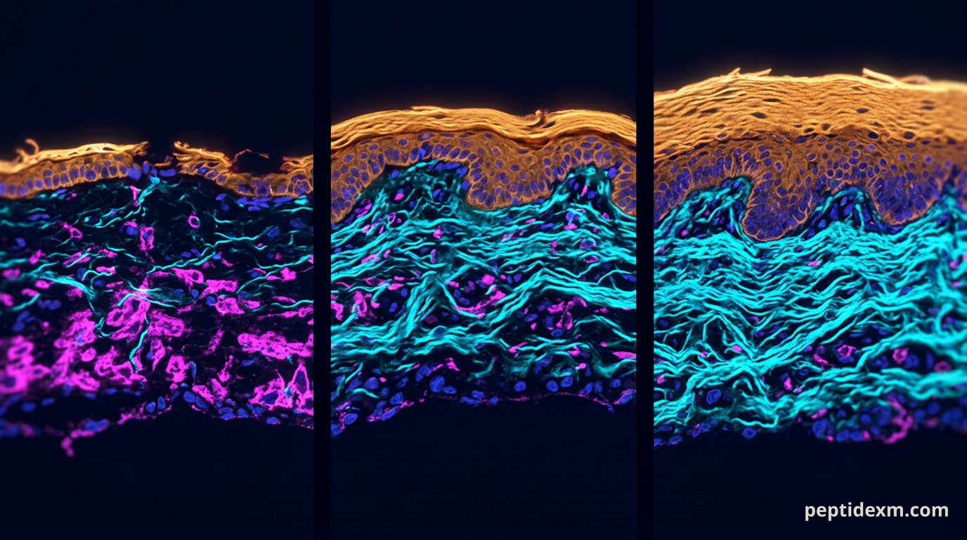
Late one winter afternoon a cosmetic scientist in Boston set three human skin explants on a bench: one treated with a copper‑binding tripeptide, one with a short growth‑factor fragment, and one with vehicle only. She tracked re‑epithelialization, collagen signal, and MMP activity over two weeks. The small dataset didn’t promise a new cosmetic blockbuster, but it taught the lab something useful about how different peptides behave in tissue — and why formulation and assay choice matter more than a label that reads “anti‑aging.”
What “peptide skincare” means in a research setting
In lab work the phrase peptide skincare refers to short amino‑acid sequences used to modulate cellular signaling, matrix turnover, inflammation markers, or pigmentation pathways in skin models. They are research reagents, not finished cosmetic products or drugs. Typical bench work ranges from simple keratinocyte or fibroblast cultures to reconstructed epidermis, ex vivo human skin explants, and small‑animal models.
Mechanisms fall into a few pragmatic categories: cell signaling modulators that bind receptors or alter second‑messenger systems; matrix modulators that influence collagen, elastin, or matrix metalloproteinases (MMPs); and peptides that modify pigmentation or barrier function. Each mechanism suggests specific readouts and controls. Don’t treat “peptide” as a single category — each sequence behaves differently in stability, uptake, and downstream biology.
Common peptide classes used in skin research
Several peptide types recur across literature and product testing. Below are the groups researchers most often evaluate, with short notes on expected lab assays:
- Copper‑binding tripeptides — often tested for effects on collagen synthesis, fibroblast migration, and wound closure assays.
- Growth‑hormone fragments and small GH‑derived peptides — assessed for proliferation and matrix gene expression in fibroblasts and explants.
- Signal‑modifying peptides (e.g., short neurotransmitter‑mimetic sequences) — usually screened in keratinocyte differentiation and inflammatory cytokine assays.
- Palmitoylated or lipidated peptides — designed to improve skin retention and evaluated for penetration and skin distribution rather than purely for bioactivity.
- Peptides targeting pigmentation — melanogenesis assays in melanocytes, co‑culture with keratinocytes, and explant pigmentation readouts.
Which one you pick depends on the hypothesis. Often teams test two or three sequences head‑to‑head so vehicle and sample handling stay constant.
GH‑derived fragment example: PE 22–28 in bench work
PE 22–28 is one of the short peptides researchers evaluate for effects that overlap with growth‑factor signaling. In vitro work typically looks at proliferation, collagen I/III mRNA, and MMP expression in dermal fibroblasts. Because the peptide is short it is prone to proteolysis in serum‑containing media; many groups use serum‑free or low‑serum conditions for short incubations, then add protease inhibitors when necessary for endpoint stability checks.

When testing PE 22–28, plan time‑course experiments. Some endpoints (eg, immediate phosphorylation of signaling kinases) are visible within minutes, while transcriptional changes in matrix genes may take 24–72 hours. Controls should include vehicle, heat‑inactivated peptide (if feasible), and a known positive control for the pathway under study.
Copper tripeptide case study: GHK‑Cu and assay selection
GHK‑Cu (glycyl‑histidyl‑lysine complexed with copper) is one of the most cited peptides in skin research. Labs test it for fibroblast migration, collagen gene expression, antioxidant gene induction, and wound‑closure assays. It also influences metalloproteinase regulation, so combined readouts make sense.
Practical points from bench reports: GHK‑Cu binds copper tightly, which alters redox chemistry; fresh stocks and careful metal controls are important. Some assays detect peptide as a mass peak, others infer activity via downstream mRNA or protein. When you see differing results across systems, check copper background in your media and reagents first.
Delivery and formulation considerations for topical testing
Peptides rarely cross intact stratum corneum unaided. For in vitro and ex vivo tests the delivery vehicle can make or break an experiment. Researchers choose vehicles that provide consistent contact, avoid cytotoxic solvents, and mimic intended product formats when possible.
Common strategies:
- Simple aqueous buffer or PBS for cell culture work, with the peptide diluted just before application.
- Oil‑in‑water emulsions or gel vehicles for explant work; these approximate cosmetic matrices and provide occlusion that can increase penetration in ex vivo skin.
- Use of penetration enhancers (ethanol, propylene glycol) or physical methods (microneedles, tape stripping) when the goal is to study transdermal delivery rather than inherent peptide activity.
Formulation stability matters. Short peptides degrade by proteolysis and by chemical pathways such as deamidation and oxidation. Consider the following mitigation strategies:
- Store aliquots frozen and avoid repeated freeze–thaw cycles.
- Use buffered systems at pH values that limit hydrolysis for your specific peptide.
- Consider lipidation or cyclization to improve skin retention; but test biological activity after modification — potency can change.
- Consider encapsulation (liposomes, ethosomes, polymeric nanoparticles) if the experiment aims to compare delivery technologies rather than intrinsic peptide biology.
In vitro and ex vivo assays that reveal meaningful effects
Design assays so they match the mechanism you hypothesise. A single readout rarely tells the whole story; couple molecular endpoints with functional ones whenever possible.
- Gene expression: qPCR for collagen types I/III, elastin, fibronectin, and MMPs.
- Protein: ELISA or western blot for procollagen C‑propeptide, MMP activity assays, and TIMP levels.
- Cellular assays: scratch wound closure, transwell migration, proliferation assays (eg, BrdU, EdU), cytotoxicity (LDH, MTT) to rule out cell death confounding results.
- Ex vivo endpoints: histology (H&E), picrosirius red for collagen organization under polarized light, immunohistochemistry for Ki‑67 or specific matrix proteins.
- Barrier and function: transepidermal water loss (TEWL) on explants, lipid lamellae visualization, and Raman spectroscopy for biochemical changes.
Selecting relevant endpoints
- If your hypothesis concerns matrix rebuilding: prioritize procollagen assays and histological collagen organization.
- If you expect anti‑inflammatory modulation: measure IL‑1β, TNF‑α, IL‑6 and corroborate with NF‑κB activation assays.
- For pigmentation studies: use melanocyte melanin quantification, tyrosinase activity, and co‑culture models with keratinocytes for transfer dynamics.
Interpreting data: common pitfalls and essential controls
Peptide research is prone to artifacts. Vehicle effects, peptide impurities, endotoxin contamination, and assay interference all create false positives. Plan controls to catch them.
Minimal control list you should include:
- Vehicle control matched for any solvents, surfactants, or lipids.
- Negative control peptide of similar length/composition but scrambled sequence to test sequence specificity.
- Heat‑inactivated or enzymatically degraded peptide to evaluate whether observed effects require the intact sequence.
- Endotoxin testing of peptide lots when working with primary cells or explants; even low endotoxin can alter cytokine readouts.
- Concentration response and time‑course experiments; single concentration/timepoint claims are weak.
Statistically, biological replicates (distinct donors or independent culture preparations) matter more than technical replicates. If you use skin explants, report donor age, anatomical site, and time from excision — those variables often explain a lot of variance.
Regulatory, sourcing, and reproducibility best practices
Buy peptides from suppliers that provide a certificate of analysis (CoA) showing purity and identity (HPLC and mass spec). For research that aims to be repeatable, store CoAs alongside raw data and record lot numbers for all reagents that contact tissue.
- Label all work as research‑use only. Do not extrapolate to human treatment or recommend self‑administration. All procurement and experiments should comply with institutional policies and applicable laws.
- Track stability: keep aliquots, document freeze‑thaw cycles, and run stability checks where possible (HPLC/MS on stored samples).
- Document handling of metal cofactors for peptides that bind metals, and test background metal levels in media and water.
- Report negative results. They improve reproducibility and reduce wasted effort in other labs.
Where a product is intended as a cosmetic ingredient you may still need to perform additional stability, preservative efficacy, and microbial challenge testing before moving to formulation claims; those are separate workflows from the mechanistic assays described earlier.
Putting it together: an example experimental workflow
Return to the Boston bench: the scientist chose GHK‑Cu and PE 22–28 because they represent distinct mechanisms — metal‑associated matrix modulation versus a short growth‑signal fragment. Her study used three donor explants per condition, applied peptides in an oil‑in‑water gel vehicle, and measured endpoints at 24 hours, 72 hours, and 14 days.
Key elements that made the dataset interpretable:
- Matched vehicle across all conditions so any occlusive or surfactant effect was controlled for.
- Endotoxin testing of peptide lots and media.
- Time points that captured immediate signaling (phospho‑kinase blot at 30 minutes), intermediate transcriptional shifts (qPCR at 24–72 hours), and matrix changes (histology and biochemical assays at 14 days).
- Documentation of donor variance: one donor showed stronger baseline MMP expression and required a paired analysis rather than pooling.
She found GHK‑Cu reduced MMP activity relative to vehicle at later time points and increased procollagen signal in two donors. PE 22–28 increased fibroblast proliferation in vitro but produced a smaller, less consistent matrix readout in explants. The result didn’t prove a clinical effect — it did, however, clarify which peptide merited deeper formulation work in that lab’s pipeline.
Peptides for skin research are tools: the sequence gives you a probable mechanism, but formulation, delivery, assays, and controls decide whether you observe that mechanism in tissue. Back on the Boston bench, the explant tray was cleared, data files saved, and the researcher planned a follow‑up that used a liposomal vehicle and an expanded donor set. The work stayed in the lab — careful, documented, and research‑only.