Peptide Strategies for Acne Research: KPV, GHK‑Cu, and Practical Workflows
When a dermatology lab in Boston applied a KPV‑containing gel to ex vivo human skin inflamed with Cutibacterium acnes, the lead researcher watched the IL‑8 signal drop by nearly half within 24 hours. No clinical claims were made. It was a bench experiment, controlled and measured; nevertheless the result pulled the team toward a more systematic study of peptides as targeted modulators of acne biology.
-
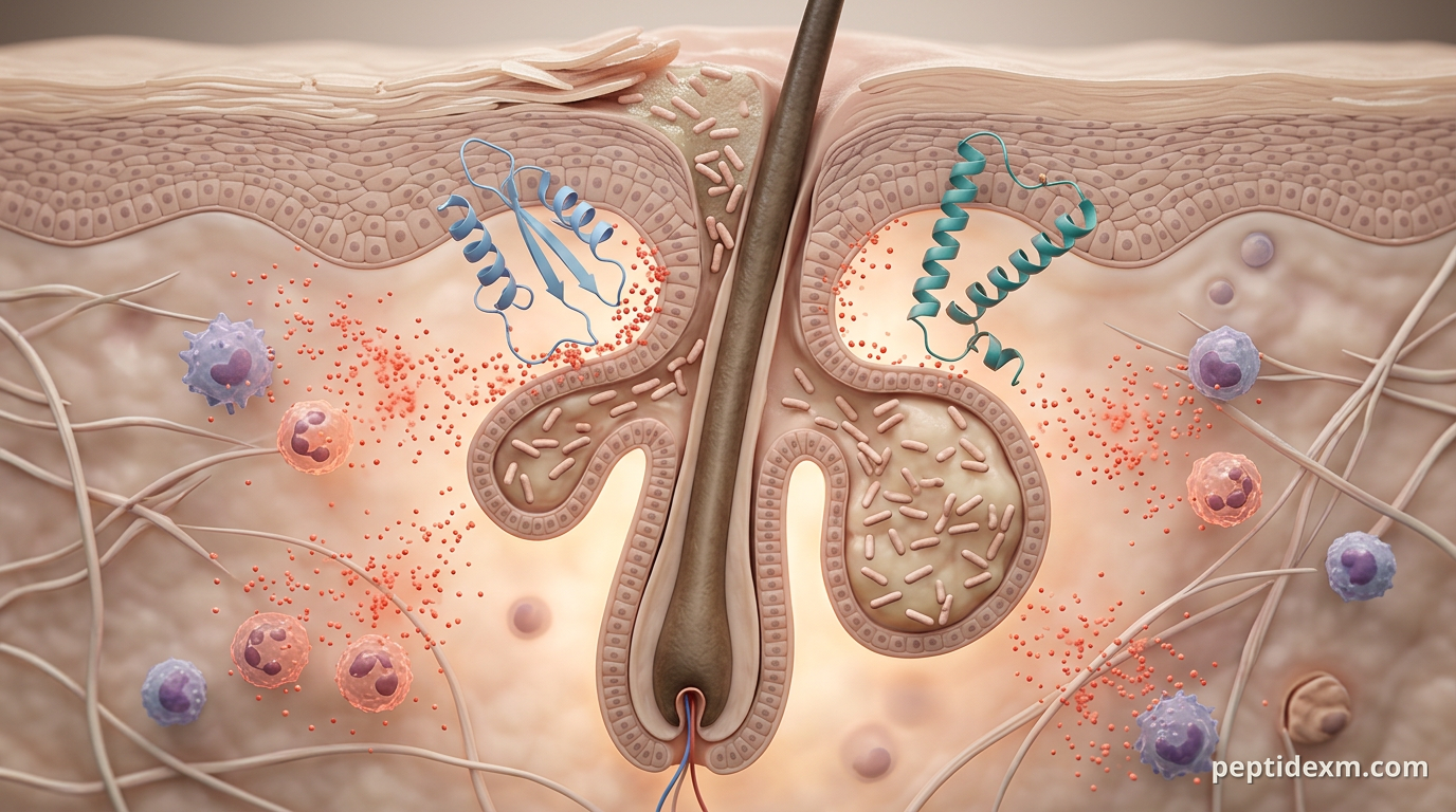
Acne pathophysiology — what a peptide can plausibly affect Research-grade acne models start with four interacting axes: sebum production, follicular hyperkeratinization, microbial communities (primarily Cutibacterium acnes), and inflammation. Each axis produces measurable molecular signals. Sebocytes secrete lipids; infundibular keratinocytes shed corneocytes that plug follicles; bacterial lipases and porphyrins trigger innate immune pathways; and inflamed tissue releases IL‑1β, IL‑6, IL‑8 (CXCL8), TNF‑α and other mediators. Peptides can act at several points in that chain. Some reduce inflammatory cytokine production in keratinocytes and sebocytes. Others enhance wound repair and collagen deposition to limit scarring. A smaller group displays direct antimicrobial or anti‑biofilm activity against C. acnes. Finally, a peptide’s effect on immune pattern‑recognition receptors (TLRs) or matrix metalloproteinases (MMPs) can change the lesion course without altering sebum output directly.
-
Why investigate peptides for acne in a research setting? Peptides bring two practical advantages to preclinical work. First, mechanism can be relatively specific: a short peptide may modulate a single receptor or enzyme without pleiotropic small‑molecule activity. Second, peptides can be chemically tuned for stability, charge, and skin penetration, which makes them good candidates for topical formulations in model systems. There are clear obstacles, too. Unmodified peptides are susceptible to proteases, they often have low permeability across the stratum corneum, and formulation components (surfactants, solvents) interact with both peptide and skin in ways that confound results. A research program should treat formulation as part of the intervention, not an afterthought.
-
Candidate peptides with preclinical evidence relevant to acne Below are peptides that show activity of potential interest to acne researchers. The summary focuses on mechanistic findings and experimental contexts, not on therapeutic recommendations.
KPV (lys‑pro‑val) — a tripeptide derived from α‑MSH that shows anti‑inflammatory and antimicrobial signatures in preclinical work. In several cell and ex vivo models KPV reduced IL‑8 and IL‑6 release from keratinocytes exposed to bacterial stimuli, and suppressed NF‑κB activation in macrophage/monocyte lines. Topical KPV formulations in rodent skin models reduced erythema and neutrophil infiltration after bacterial challenge. The peptide is small and polar, so formulation must address skin penetration and proteolysis.
GHK‑Cu (glycyl‑histidyl‑lysine‑copper) — a copper‑binding tripeptide best known for promoting collagen synthesis, reducing MMP expression, and accelerating wound closure in dermal models. In keratinocytes and fibroblasts GHK‑Cu increases collagen I and III mRNA and decreases MMP‑2 and MMP‑9 activity; it also has demonstrable anti‑inflammatory effects, such as lowering TNF‑α and IL‑6 in some in vitro systems. Those properties make GHK‑Cu appealing for post‑inflammatory remodeling and scar attenuation after inflammatory lesions.
BPC‑157 — a gastric peptide fragment that has been studied primarily for wound healing and tissue repair. Preclinical studies show accelerated angiogenesis and granulation tissue formation and modulation of inflammatory cytokines. In skin wound models BPC‑157 improves closure kinetics and may alter collagen deposition patterns, which is useful information for research into scar formation after acne lesions.
Thymosin beta‑4 (TB‑500) — promotes cell migration and matrix remodeling through actin modulation and can speed re‑epithelialization in wound models. It reduces inflammatory cell infiltration in some settings, which explains interest for studies on lesion resolution and scar prevention.
Thymosin alpha‑1 — an immunomodulatory peptide that alters TLR signaling and cytokine profiles in immune cells. Preclinical data indicate it can shift innate immune responses without global immunosuppression, a property that may help in models where dampening overactive inflammation rather than killing bacteria is the experimental goal.
Synthetic antimicrobial peptides (AMPs) — a broad category that includes modified defensins, protegrin analogs and novel sequences designed to disrupt bacterial membranes or biofilms. Several synthetic AMPs show MICs against C. acnes in vitro and prevent biofilm formation on ex vivo follicular models, but toxicity to keratinocytes is a critical experimental endpoint.
- Formulation and delivery — practical constraints for skin work Topical delivery is the dominant route in acne research because it minimizes systemic exposure and lets you test local mechanisms. Still, delivering an intact peptide to the hair follicle is nontrivial. The stratum corneum limits diffusion of polar peptides; follicular targeting requires either penetration enhancers, lipid carriers, or physical methods (microneedling, tape stripping) in controlled experiments. Common formulation strategies in preclinical studies
Liposomes and niosomes to protect peptides from proteases and improve follicular uptake. Hydrogel bases (carbomers, hyaluronic acid) for sustained local release and reduced irritation in explant models. Nanoparticle encapsulation (PLGA, chitosan) to control release kinetics and favor follicular retention. Permeation enhancers (e.g., low‑percent ethanol, propylene glycol) used with caution because they change skin barrier properties and inflammatory readouts.
- Assays and experimental workflows for acne peptide studies Designing an experiment requires clear primary endpoints. Typical endpoints are cytokine production (IL‑1β, IL‑6, IL‑8), sebum lipid profile, bacterial counts/biofilm biomass, histologic inflammation score, and collagen/matrix remodeling markers for scarring studies.
Short protocol outline (in vitro → ex vivo → in vivo)
In vitro: treat immortalized human sebocytes (e.g., SEB‑1) and keratinocytes (HaCaT or primary cells) with peptide ± C. acnes lysate; measure IL‑8/IL‑6 by ELISA at 6, 24, 48 hours. Co‑culture: establish keratinocyte–sebocyte co‑culture with live or heat‑killed C. acnes to assess peptide effects on cytokine crosstalk and lipid secretion. Biofilm and MIC: perform planktonic MIC assays and crystal violet biofilm assays against reference C. acnes strains; include keratinocyte cytotoxicity assays (MTT, LDH) to define therapeutic index in vitro. Ex vivo human skin explants: apply formulated peptide to follicular units or tape‑stripped skin, then challenge with C. acnes; sample tissue for qPCR, ELISA, histology at 24–72 hours. Small‑animal models: if ethically approved, use well‑characterised acne models (e.g., mouse ear injection or intradermal C. acnes) to assess inflammatory lesion size, immune cell infiltration, and scarring endpoints over days to weeks.
Key readouts and assays
Cytokines: ELISA or multiplex panels for IL‑1β, IL‑6, IL‑8, TNF‑α, MCP‑1. Gene expression: qPCR for TLR2, MMPs, collagens, and inflammatory transcription factors (NF‑κB components). Bacterial load: CFU counts from homogenized tissue or qPCR for bacterial DNA; biofilm biomass by crystal violet. Lipidomics: GC‑MS or LC‑MS to quantify sebum triglycerides, wax esters and free fatty acids. Histology and immunohistochemistry: neutrophil markers (MPO), macrophage markers (F4/80 in mouse), collagen I/III staining for remodeling.
- Typical experimental parameters and controls In vitro concentrations for peptides commonly range from 0.1 to 100 µM, depending on potency and cytotoxicity. Start with a broad range (0.1, 1, 10, 50 µM) and include vehicle controls and a positive comparator if available (for example, an anti‑inflammatory peptide with established activity). Timepoints normally include acute (6–24 h for cytokines) and subacute (48–72 h for matrix changes). Essential controls
Vehicle alone (identical formulation without peptide). Peptide scrambled sequence to control for sequence‑specific effects. Endotoxin testing and matched endotoxin controls — bacterial products can masquerade as peptide effects. Keratinocyte and sebocyte viability controls to separate cytotoxicity from anti‑inflammatory activity.
-
Common pitfalls and how to address them Proteolysis and loss of effective concentration are frequent. Include protease inhibitors in initial in vitro screens to determine whether loss of activity is due to degradation. For topical studies, check peptide integrity after 24–72 hours in the formulation and on skin explants using LC‑MS. Another common confounder is formulation interference with assays. Surfactants or solvents used to solubilize peptides can directly alter cytokine release or cell viability. When possible, choose formulations that have been validated in the same cell types and run parallel vehicle assays. Batch variability matters. Synthesis impurities, truncated sequences, and copper content (for GHK‑Cu) change activity dramatically. Always analyse peptide lots by HPLC and mass spectrometry and report lot numbers in methods.
-
Regulatory, safety, and ethical context for research use All work described here is preclinical. Peptides supplied for research must be labeled and used as research reagents only, not for human administration. Institutional approvals (IACUC for animal work, IRB/ESC for human explant use) are required where applicable. For topical or animal studies consider sterility, endotoxin limits, and GLP standards if the project aims to generate regulatory filings. Safety screening in early stages should include standard toxicology assays: local irritation, sensitization panels, and systemic exposure checks if systemic absorption is plausible. Record and report any adverse effects in animals following institutional guidelines.
-
Case study: translating a bench observation into a structured study Return to the lab hook. The Boston team that observed the IL‑8 drop with a KPV gel designed a follow‑up study with these steps: (1) reproduce the IL‑8 result in primary keratinocytes and sebocytes at three peptide concentrations; (2) run C. acnes biofilm assays to see whether KPV has direct antimicrobial activity; (3) test a KPV+GHK‑Cu combination in ex vivo human skin explants to assess both inflammation and early matrix remodeling; (4) verify peptide integrity on skin after 24 hours by LC‑MS; (5) include scrambled‑KPV controls and endotoxin assays. Their initial data were cautious but consistent: KPV lowered IL‑8 and IL‑6 in keratinocytes exposed to bacterial stimuli, while GHK‑Cu increased pro‑collagen mRNA in fibroblasts in the same explants. Combination treatment did not increase keratinocyte toxicity. Those results justified a deeper timecourse and mechanistic assays (NF‑κB nuclear translocation, MMP activity assays) before any in vivo animal work under an approved protocol.
For researchers, the practical takeaway is simple: peptides offer targeted biological handles on the inflammation and repair phases of acne, but success depends on rigorous formulation work, appropriate controls, and transparent reporting of lot‑to‑lot and stability data.
Back in the Boston lab the researcher closed the incubator, labeled the samples, and wrote the methods section with a clearer plan. The IL‑8 trace on the ELISA sheet was still the same — a small, repeatable signal pointing to a set of experiments worth doing, strictly in the lab and strictly for research purposes.