How Peptides Kill Cancer Cells: Mechanisms, Examples, and Research Considerations
Peptides are small enough to reach intracellular targets and specific enough to avoid some off-target effects of small molecules. That combination has made them a fast-growing tool in cancer research — from cell-killing membranolytic sequences to peptide vaccines and delivery tags that ferry drugs into tumors. This post gives you a compact, practical tour: mechanisms, representative peptide classes, common assays, and the logistical details you’ll actually use in the lab.
Why researchers look to peptides in cancer work Peptides sit between small molecules and antibodies. They’re typically 5–50 amino acids long, so they can engage protein surfaces that small molecules miss but are much cheaper and easier to make than monoclonal antibodies. You can synthesize them with high purity, add chemical stabilizers, or stitch them onto carriers. That flexibility is why labs exploring targeted cytotoxicity, intracellular disruption of protein complexes, or immune modulation often reach for peptides. Put another way: if you need something modular, tunable, and sequence-driven — and you’re working at the bench rather than in large-scale GMP manufacturing — peptides are a pragmatic choice. The trade-offs are stability, delivery, and sometimes selectivity. Those are solvable, but they change the experiments you design.
Major mechanisms by which peptides kill or suppress tumor cells Peptides can affect tumors through several distinct mechanisms. Each has its own assay set, failure modes, and design levers. Knowing which mechanism you want to test will save you time and money.
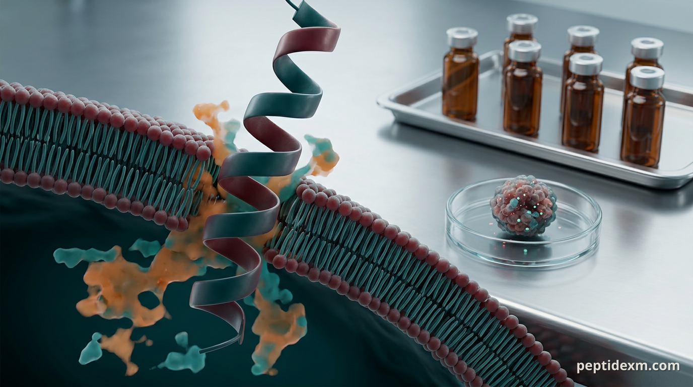
Membrane-active peptides (AMPs) Many anticancer peptides are cousins of antimicrobial peptides. They disrupt membranes directly, forming pores or destabilizing bilayers rich in anionic lipids. Tumor cells often expose more negatively charged phospholipids on their outer leaflet and have higher membrane potential, which can make them relatively susceptible to cationic, amphipathic peptides.
Strength: rapid cytotoxicity visible in short-term viability assays. Weakness: hemolysis and toxicity to non-tumor tissues if systemic exposure is high. Typical readouts: lactate dehydrogenase (LDH) release, membrane-impermeant dyes, and short incubation live/dead assays.
Apoptosis induction and PPI disruption Some peptides bind intracellular proteins and trigger programmed cell death. BH3-mimetic peptides, stapled peptides that stabilize helical structures, and fragments that block protein–protein interactions (for example, p53–MDM2) fall into this category. These require delivery across the plasma membrane or fusion to cell-penetrating sequences.
Targeted receptor binding and receptor-mediated internalization Peptides can be designed to bind tumor-associated receptors and either trigger death pathways or act as a Trojan horse to deliver cargo (toxins, radionuclides, nanoparticles). RGD motifs that bind integrins and tumor-penetrating sequences like iRGD are common tools.
Immune modulation and peptide vaccines Short peptides representing tumor antigens are used to prime T cells in vitro and in vivo. Other immunomodulatory peptides tune innate responses or act as adjuvants. These approaches don’t directly lyse cancer cells but can produce cytotoxic T-cell responses that do.
Angiogenesis inhibition and microenvironment effects Some peptides interfere with new vessel growth or with stromal interactions necessary for tumor survival. They can be fragments of larger extracellular-matrix proteins or engineered sequences that compete with pro-angiogenic factors.
Representative peptide classes and research examples Below are commonly studied peptide types, with the experimental contexts where they typically show effect. I focus on research evidence — cell lines, spheroids, and animal models — not clinical claims.
Melittin and membranolytic peptides. Melittin, from bee venom, rapidly lyses membranes in vitro and shrinks tumors in murine models when delivered locally or packaged in nanoparticles to limit systemic toxicity. Lots of papers use melittin-derived sequences to test membrane-targeted strategies; keep an eye on hemolysis assays. Lactoferricin and defensin-derived peptides. These host-defense fragments show selective cytotoxicity in several cancer cell lines and reduce tumor growth in xenograft work. Mechanisms mix membrane disruption and intracellular signaling perturbation. Stapled peptides and BH3 mimetics. Stapling stabilizes α-helices and improves cell penetration and protease resistance. Researchers use stapled BH3 peptides to antagonize BCL-2 family proteins and trigger apoptosis in apoptosis-resistant lines. p53-derived peptides and MDM2 inhibitors. Short peptides that reconstitute p53 activity or block its negative regulators can restore apoptosis pathways in p53-wildtype models or sensitize cells to chemotherapy in combination studies. Tumor-penetrating peptides (iRGD, RGD variants). These aren’t usually cytotoxic alone; instead, they enhance delivery of chemotherapeutics or nanoparticles into tumors in murine models and 3D spheroid systems. Immunomodulatory peptides. Thymic peptides, certain TLR‑agonist peptides, and tumor antigen peptides are widely used as adjuvants or vaccine candidates in preclinical immuno-oncology studies.
Tumor targeting and delivery strategies Delivering a peptide to a tumor is where many projects live or die. You can design a peptide that’s active in vitro and completely inert in vivo if it never reaches the tumor at sufficient concentration. Common approaches reduce that risk.
Conjugation to tumor-homing motifs (e.g., RGD, iRGD) to exploit receptor-mediated uptake. Packaging in liposomes, polymer nanoparticles, or cell-derived vesicles to improve circulation half-life and reduce off-target interactions. Chemical stabilization: cyclization, D-amino acids, N-methylation, stapling, or PEGylation to boost protease resistance. Prodrug strategies where the peptide is activated by tumor-enriched proteases (e.g., MMPs).
For immune-focused peptides, intradermal or subcutaneous delivery is commonly used in animal models to prime T cells. For direct-cytotoxic peptides, local delivery or nanoparticle encapsulation frequently improves therapeutic index in mouse models.
Practical assays and models you’ll likely run Pick your assays based on mechanism. If you suspect membrane lysis, short-term LDH or PI uptake assays are fast and cheap. If you expect apoptosis from PPI disruption, caspase activity, Annexin V, and time-course viability assays are more informative. A few other common tools:
2D cytotoxicity assays (MTT/XTT/CellTiter-Glo) for initial screening. 3D spheroids and organoids to test penetration and activity in a more tissue-like context. Co-culture systems with immune cells if you’re testing antigenic peptides or immune modulators. Mouse xenograft or syngeneic tumor models for in vivo efficacy and biodistribution. Biodistribution imaging (fluorescent or radiolabeled peptides) and LC-MS for PK/PD measurements.
Design early experiments to answer the key translational questions: does the peptide reach the tumor? Does it engage the intended molecular target? Is toxicity tolerable in the intended delivery format?
Common challenges and how research teams handle them Peptide projects tend to hit the same friction points. Being explicit about them saves months.
Proteolysis. Peptides are substrates for serum and tissue proteases. You can cyclize, substitute D-amino acids, or use stapling to increase half-life in plasma. Each modification can affect activity, so re-test functional assays after modification. Off-target membrane toxicity. Membranolytic peptides can cause hemolysis. Standard counter-screens include human red blood cell assays and primary cell cytotoxicity panels. Delivery to solid tumors. Dense stroma and high interstitial pressure block penetration. Tumor-penetrating motifs and nanoparticle carriers are commonly explored to address that. Immunogenicity. Peptides can provoke antibody responses that clear them from circulation. Humanized sequences or shielding chemistries reduce this risk but can complicate synthesis. Quantitation in vivo. Small peptides clear quickly, so sensitive LC-MS or radiolabeling is often necessary to measure exposure.
Regulatory, quality, and translational considerations for lab procurement Even at the research stage, quality matters. Peptide impurities and endotoxin can confound results — especially in immunology experiments. Look for vendors that provide:
Analytical HPLC and mass-spectrometry purity reports. Endotoxin testing if you’ll use the peptide in immune assays or in vivo work. Certificates of analysis that list lot-specific data and recommended storage.
And remember: research peptides are for laboratory use only. They aren’t approved therapeutics. Protocols that move toward human testing require GMP-grade material, regulatory approvals, and different manufacturing workflows.
Practical sourcing and handling tips Little details trip up experiments more often than big conceptual errors. A few practical pointers:
Store lyophilized peptides at −20 °C or colder, in a dry environment. Avoid repeated freeze–thaw cycles on reconstituted material. Hydrophobic peptides often need organic solvents like DMSO for complete dissolution before dilution into aqueous buffers. Check solubility notes in the COA. Confirm reconstitution solvent is compatible with downstream assays and cells. Even low DMSO percentages can change membrane behavior in short-term assays. Request endotoxin testing for peptides destined for cell-culture experiments involving immune readouts or for in vivo administration.


Outlook: where peptide-based cancer research is headed Two trends stand out. First, hybrid approaches where peptides act as targeting modules for larger therapeutic payloads — ADC-like constructs with peptide homing sequences — are proliferating in preclinical work. Second, chemical stabilization (stapling, cyclization, noncanonical residues) plus smart delivery systems is making previously fragile intracellular-targeting peptides experimentally tractable. If you work with peptides in tumor models, expect the iterative cycle to look something like: design → in vitro mechanism assays → 3D penetration testing → PK/biodistribution → targeted in vivo proof-of-concept. Each step answers a distinct decision point about chemistry, formulation, and delivery.
Peptides won’t replace small molecules or antibodies overnight. But as tools for probing tumor biology and as modular payloads for targeted delivery, they’re here to stay. Use robust in vitro-to-in vivo testing, insist on clean analytical data, and match your peptide chemistry to the biological question you’re asking — and you’ll avoid the common dead-ends most labs run into.