Best Peptides for Skin and Beauty: A Research Guide

Peptides are a common tool in skin-research protocols. They target fibroblasts, keratinocytes, melanocytes, and extracellular matrix turnover with a degree of specificity small molecules rarely match. This guide compares leading skin-focused peptides, shows how to design studies around them, and highlights the delivery and measurement choices that matter most. All material is for research use only.
Why peptides for skin research? Peptides are short chains of amino acids that act as signaling molecules. In skin, they can modulate collagen synthesis, inflammation, pigmentation, and wound repair. Their small size helps with tissue penetration, and you can sequence or modify them to alter stability and receptor selectivity. For labs, that means targeted mechanistic experiments are feasible without the confounding effects of large proteins. Concrete takeaway: pick peptides when you need pathway-level modulation (for example, stimulating collagen I expression or altering MMP activity) rather than broad cytotoxic effects. They lend themselves to dose–response curves, time-course analysis, and combination testing with topical vehicles or physical penetration aids.
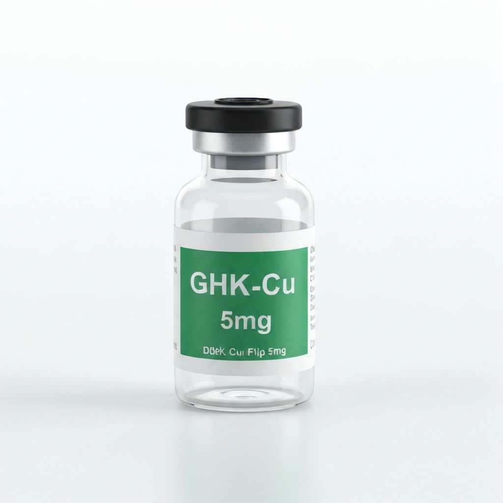
GHK‑Cu (copper tripeptide): what it does and how to study it GHK‑Cu (glycyl‑histidyl‑lysine complexed with copper) is a well-characterized peptide for skin research. It interacts with metalloproteinases, stimulates collagen and glycosaminoglycan synthesis in fibroblasts, and has been reported to influence wound-healing gene networks in transcriptomic studies. It also binds copper, which can facilitate redox chemistry—an aspect worth monitoring in vitro.
When to use it Use GHK‑Cu when the endpoint is matrix remodeling, collagen deposition, or keratinocyte migration. It's a frequent choice for wound-healing models and skin-equivalent cultures where extracellular matrix readouts are primary.
How a study might design this Start with dose–response in primary human dermal fibroblasts: 0.1–1000 nM is a common in vitro range, though precise concentrations depend on the assay sensitivity. Include timepoints at 24, 48, and 72 hours for gene expression and up to 7–14 days for collagen deposition assays. Consider co-culture with keratinocytes for epidermal–dermal signaling.
What to avoid Don’t assume stability in every vehicle. GHK‑Cu can dissociate copper under harsh oxidizing conditions; verify peptide integrity by LC‑MS if your formulation includes pro-oxidant components. Also avoid single-endpoint measurements; MMP transcripts can transiently rise even while net collagen increases. The concrete control: always pair transcriptional and protein-level assays.
SNAP‑8 (snap-8): a peptide for expression-level changes in contraction and wrinkles SNAP‑8 is an octapeptide derived from SNAP‑25 sequences that modulates neurotransmitter release pathways in the skin’s neuromuscular-like units. In topical research it’s often evaluated for its capacity to reduce contraction-related microfolds in ex vivo skin and full-thickness constructs. Mechanistically, it interferes with SNARE complex formation, which can alter muscle-associated contraction around follicles.
When to use it Pick SNAP‑8 for ex vivo skin explants, reconstructed skin models, or organotypic cultures where microcontraction or pore/tension-related endpoints are measurable. It’s not a collagen stimulator; it’s about reducing surface contraction and dynamic wrinkle depth in imaging assays.
How a study might design this Apply SNAP‑8 topically in a defined vehicle across an explant panel. Use high-resolution 3D profilometry or optical coherence tomography to quantify surface geometry before and after treatment, with at least three timepoints across 1–4 weeks. Add physiological contraction controls—e.g., electrical field stimulation if using innervated models—to stress the system and observe peptide efficacy under load.
What to avoid Avoid relying on subjective scoring. Surface-imaging metrics are sensitive to lighting and handling; standardize imaging conditions and use automated analysis where possible. If your preparation includes proteases (wounds, inflamed skin), protect SNAP‑8 from rapid degradation or quantify intact peptide over time.
PE 22–28: collagen induction and matrix remodeling PE 22–28 is a peptide fragment studied for its effect on collagen turnover and elastin expression. It’s often tested in models focusing on dermal thickness, tensile strength, and histologic matrix organization. The mechanism appears to be fibroblast-mediated remodeling rather than direct scaffolding.
When to use it Use PE 22–28 when dermal architecture changes are the primary outcome. It's suited to animal skin models, ex vivo human dermis, and engineered dermal matrices where tensile testing and histomorphometry are planned.
How a study might design this Combine histology (Masson’s trichrome for collagen), hydroxyproline assays for total collagen content, and second-harmonic generation (SHG) imaging to evaluate fiber organization. Timeframes are typically longer than for transcriptional readouts—expect measurable changes at 2–8 weeks in organ or animal models. Pair with mechanical testing for functional readouts.
What to avoid Do not equate increased collagen quantity with improved skin mechanics. Disorganized collagen can increase bulk but reduce tensile strength. Always include organization metrics (SHG or polarized light microscopy) and at least one functional assay.
Melanotan‑2: pigmentation research and safety considerations Melanotan‑2 is commonly used in pigmentation studies because it acts on melanocortin receptors that regulate melanogenesis. In controlled models it’s useful for studying melanocyte activation, pigment transfer to keratinocytes, and signaling downstream of MC1R pathways.
When to use it Select Melanotan‑2 when pigment production, distribution, or UV-response modulation are your endpoints. It’s appropriate in melanocyte cultures, reconstructed epidermis, and small-animal pigment models.
How a study might design this Measure tyrosinase activity, melanin content, and melanosome transfer assays. Include UV-exposed and non-exposed arms to assess interaction with DNA-damage repair pathways. Timepoints often run from 48 hours (enzyme activity) to 7–14 days (visible pigment changes in tissue models).
What to avoid Avoid oversimplified pigmentation readouts. Spectrophotometric melanin measurements can conflate tissue absorption and scattering. Use chemical quantification (e.g., NaOH solubilization and absorbance) plus imaging-based localization. Also plan for appropriate biosafety handling; receptor activation can have wide downstream effects in vivo.
Blends and combination approaches: when multi-target makes sense Single peptides hit specific nodes. Blends can target multiple nodes—matrix synthesis, contraction, pigmentation, and inflammation—simultaneously. This often improves effect size in complex tissue models but complicates mechanistic attribution.
When to use blends Use blends when your outcome is a composite skin benefit (for example, reduced wrinkle depth plus improved hydration). They’re useful in translational explant studies where the goal is proof of overall tissue improvement rather than mechanistic dissection.
How a study might design this Design factorial experiments. Test individual components and their combinations to identify synergy or antagonism. Include at least one readout per target pathway—collagen assays, contraction imaging, pigment quantification—to map which components drive which outcomes.
What to avoid Don’t run a blend without component controls. A positive result from a blend is uninterpretable unless each peptide’s contribution is known. Consider starting with a two-by-two design for practical throughput.
Topical versus systemic delivery: stability, vehicle, and penetration Delivery determines whether the peptide reaches its target cell type. For skin research, topical is the default, but barriers remain. Stratum corneum limits macromolecule passage. Formulation choices—oligopeptides, penetration enhancers, encapsulation, or microneedle application—change bioavailability dramatically.
When to choose topical, intradermal, or systemic Topical: choose when target cells are epidermal or superficial dermal and when maintaining a localized exposure is desired. Intradermal injection: useful for direct dermal fibroblast or melanocyte targeting and for avoiding stratum corneum limitations. Systemic: rarely required for skin endpoints unless you study endocrine interactions or immune trafficking that require systemic exposure.
How a study might design delivery arms Include a vehicle-only control. If testing topical penetration, pair with tape-stripping or Franz diffusion cells and quantify intact peptide in receptor fluid by LC‑MS. For in vivo or ex vivo intradermal delivery, standardize injection volume and depth and measure local concentration-time profiles.
What to avoid Don’t assume a peptide is stable in your vehicle. Aqueous gels, solvent blends, and emulsions can hydrolyze peptides or cause aggregation. Test stability at experimental temperatures and durations, and verify bioactivity after formulation where possible.
Endpoint selection and assays Choose endpoints that measure mechanism and function. Histology and gene expression alone don’t prove functional improvement. Combine molecular, structural, and biomechanical assays for a credible story.
Quantitative imaging techniques High-value imaging includes second-harmonic generation (SHG) for collagen organization, confocal microscopy for melanosome transfer, optical coherence tomography (OCT) for live tissue geometry, and 3D profilometry for surface topology. These techniques reduce observer bias and provide spatial context that biochemical assays lack.
Gene/protein: qPCR for collagen I/III, MMPs, TIMPs; ELISA or western blot for secreted markers. Matrix: Hydroxyproline for total collagen, SHG for fiber organization, polarized light microscopy for birefringence. Function: Tensile testing, TEWL (transepidermal water loss) for barrier integrity, contractility assays for explants.
Concrete takeaway: include at least one functional assay (mechanical test, TEWL, or live imaging) in every protocol aimed at demonstrating a meaningful tissue change.
Common experimental pitfalls and controls Peptide work is deceptively simple. Small mistakes cause big misinterpretation. Here are the recurring problems labs report and how to fix them.
Stability blind spots: Test peptide integrity under actual experimental conditions (vehicle, temperature, light). LC‑MS or HPLC is cheap insurance. Delivery assumptions: Confirm delivery with tracer assays or direct peptide quantification in tissue, especially for topicals. Single-readout traps: Don’t rely on one assay. Pair transcriptional data with protein and functional measurements. Batch variability: Biological materials (primary cells, explants) vary. Use multiple donors and report donor numbers; randomize treatment assignments.
Concrete takeaway: pre-define primary and secondary endpoints, run stability and delivery checks before committing to long experiments, and include adequate biological replicates.
Practical checklist before you run an experiment Before starting: confirm peptide identity and purity with analytical data, verify stability in your vehicle for the planned exposure time, and perform a brief pilot to set concentration ranges and timepoints. Decide on at least one functional endpoint and the minimal number of biological replicates needed to detect expected effect sizes.
Confirm LC‑MS/HPLC identity and purity. Run 24–72 hour stability in vehicle at assay temperature. Do a 3‑point pilot for dose and choose at least three timepoints. Include vehicle, positive control (where appropriate), and at least three biological replicates per arm.
Final takeaway: peptides give specific, tunable ways to change skin biology in research settings. The key to useful results is thoughtful design—validate delivery, pair mechanisms with function, and control for stability and biological variability. This keeps your data interpretable and reproducible.